Epithelial Tissue
Epithelial Tissue
Epithelial tissue is a sheet of cells that covers a body surface or lines a body cavity. Two forms occur in the human body:
- Covering and lining epithelium– forms the outer layer of the skin; lines open cavities of the digestive and respiratory systems; covers the walls of organs of the closed ventral body cavity.
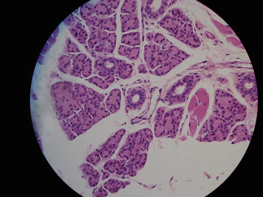
- Glandular epithelium– surrounds glands within the body.
Characteristics of epithelium
Epithelial tissues have five main characteristics.
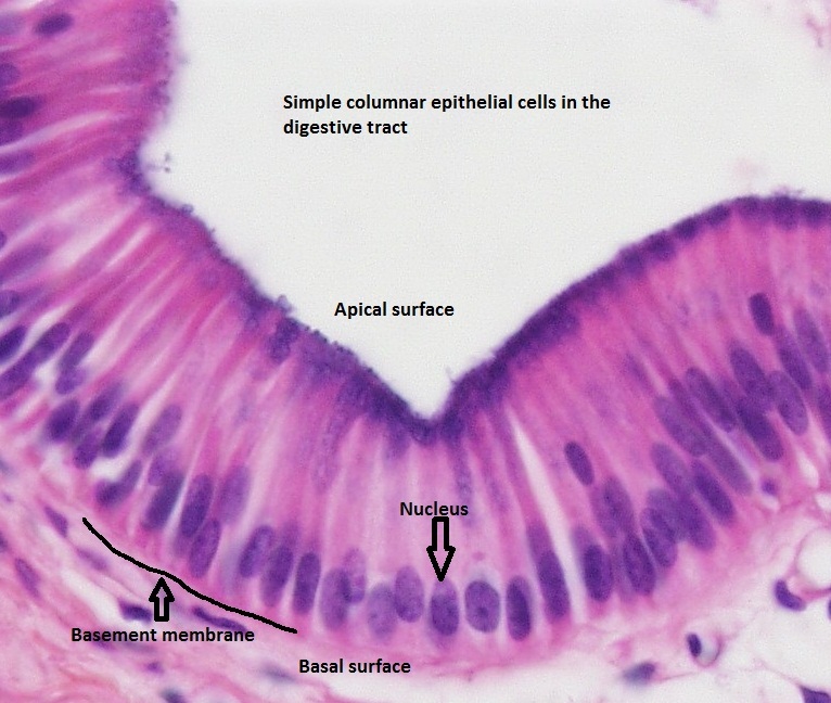
- Polarity– all epithelia have an apical surface and a lower attached basal surface that differ in structure and function. For this reason, epithelia is described as exhibiting apical basal polarity. Most apical surfaces have microvilli (small extensions of the plasma membrane) that increase surface area. For instance, in epithelia that absorb or secrete substances, the microvilli are extremely dense giving the cells a fuz
 zy appearance called a brush border. Examples of this would include epithelia lining the intestine and kidney tubules. Other epithelia have motile cilia (hairlike projections) that push substances along their free surface. Next to the basal surface is the basal lamina (thin supporting sheet). The basal lamina acts as a filter allowing and inhibiting certain molecules from passing into the epithelium.
 - Specialized contacts– epithelial cells fit close together and form continuous sheets (except in the case of glandular epithelia). They do this with tight junctions and desmosomes. Tight junctions form the closest contact between cells and help keep proteins in the apical region of the plasma membrane. Desmosomes connect the plasma membrane to intermediate filaments in the cytoplasm.
- Supported by connective tissue– all epithelia are supported by connective tissue. For instance, deep to the basal lamina is reticular lamina (extracellular material containing collagen protein fiber) which forms the basement membrane. The basement membrane reinforces the epithelium and helps it resist stretching and tearing.
- Avascular and innervated– even though epithelium is avascular (contains no blood vessels), it’s still innervated (supplied by nerve fibers).
- Regeneration– epithelium have a high regenerative capacity and can reproduce rapidly as long as they receive adequate nutrition.
Classification of Epithelia
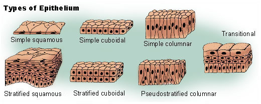
Epithelium has two names. The first name indicates the number of cell layers, the second describes the shape of its cell. Based on the number of cell layers, epithelia can either be simple or stratified.
- Simple epithelia– consist of a single cell layer (found where absorption, secretion, and filtration occur).
- Stratified epithelia– are composed of two or more cell layers stacked on top of each other (typically found in high abrasion areas where protection is needed).
All epithelial cells have six sides but they vary in height. For this reason, there are three ways to describe the shape and height of epithelial cells.
- Squamous cells– are flat and scale-like.
- Cuboidal cells– are box-like (same height and width).
- Columnar cells– are tall (column shaped).
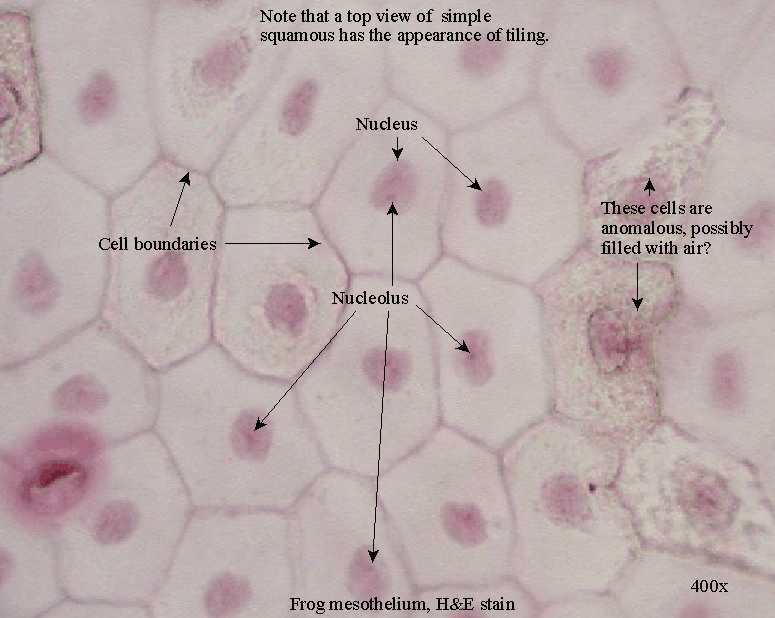
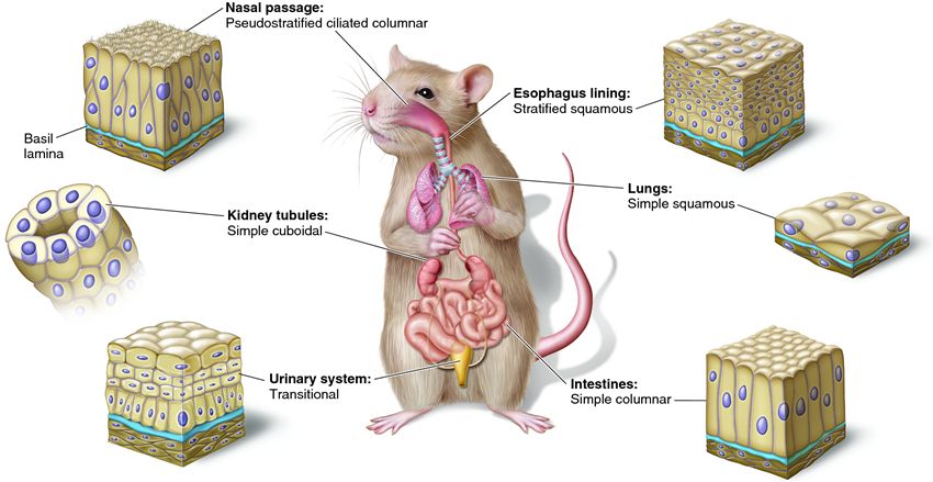
Simple squamous epithelium– are close fitting and flattened laterally. They’re found where filtration occurs (kidneys, lungs) and they resemble the look of a fried egg. Two simple squamous epithelia in the body have special names reflecting their location.
- Endothelium– provides a friction-reducing ling in lymphatic vessels and all hollow organs of the cardiovascular system (heart, blood vessels, capillaries).
- Mesothelium– is the epithelium found in serous membranes (membranes lining the ventral body cavity and covering the organs within it).
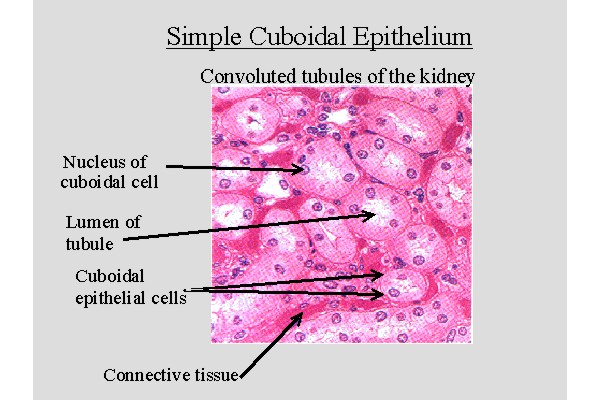
Simple cuboidal epithelium– consists of a single layer of cells with the same height and width. Functions include secretion and absorption (located in small ducts of glands and kidney tubules).
Simple columnar epithelium– is a single layer of tall, closely packed cells that line the digestive tract from the stomach to the rectum. Functions include absorption and secretion. They contain dense microvilli on their apical surface . Additionally, some simple columnar epithelia may display cilia on their free surface also.
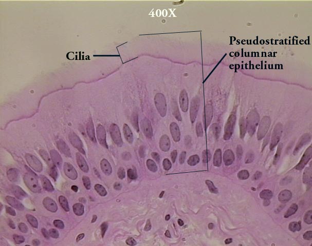
Pseudostratified columnar epithelium– vary in height. All of their cells rest on the basement membrane and only the tallest reach the apical surface. When viewing pseudostratified epithelium it may look like there are several layers of cells, but this is not the case. (because the cells have different heights, it gives the illusion of multiple cell layers). Most pseudostratified epithelia contain cilia on their apical surface and line the respiratory tract.
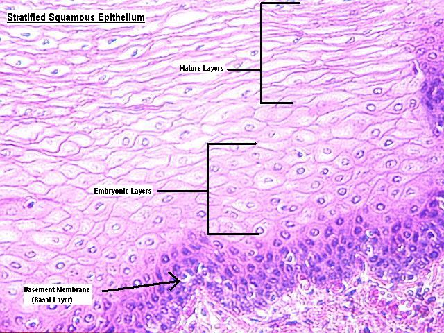
Stratified squamous epithelium– is the most widespread stratified epithelia. It’s composed of several layers and is perfect for its protective role. Its apical surface cells are squamous and cells of the deeper layer are either cuboidal or columnar. Stratified squamous forms the external part of the skin and extends into every body opening that’s continuous with the skin. The outer layer of the skin (epidermis) is keratinized (contains keratin, a protective protein). Other stratified squamous in the body is nonkeratinized.
Stratified cuboidal epithelium
Stratified cuboidal epithelium– is somewhat rare in the human body. It’s mainly found in the ducts of glands (sweat glands, mammary glands) and is typically has two layers of cuboidal cells.
Stratified columnar epithelium
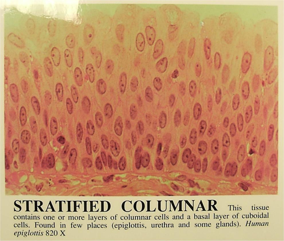
Stratified columnar epithelium– is also rare in the human body. Small amounts are found in the pharynx, male urethra, and lining of some glandular ducts. Stratified columnar epithelium occurs in transition areas (junctions) between other epithial types.
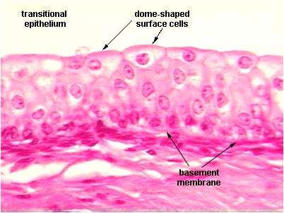
Transitional epithelium– forms the lining of hollow urinary organs, which stretch as they fill with urine. Cells in the basal layer are cuboidal or columnar. Cells by the apical surface vary in appearance depending if the organ is stretched at the time. Transitional cells have the ability to change their shape which allows more urine to flow through.
For information on glandular epithelium, click here.
Related Posts
Category: Tissue

