Body cavities and membranes
Body cavities and membranes
In most cases, the body is described as having two main cavities called the “dorsal and ventral body cavities”. Some anatomical references do not recognize the dorsal body cavity but we will use it in this example because it’s used by many professionals and colleges.
It also makes it easier to understand! If you would like to brush up on anatomical language and directional terms before moving on, click here.
Dorsal body cavity
The dorsal body cavity protects organs of the nervous system and has two subdivisions. The cranial cavity is the area within the skull and encloses the brain. The spinal (vertebral) cavity encases the vertebral column and spinal cord.
Ventral Body cavity
Like the dorsal cavity, the ventral cavity has two subdivisions. The superior division is called the thoracic cavity. The thoracic cavity is surrounded by the ribs and muscles in the chest. It’s further su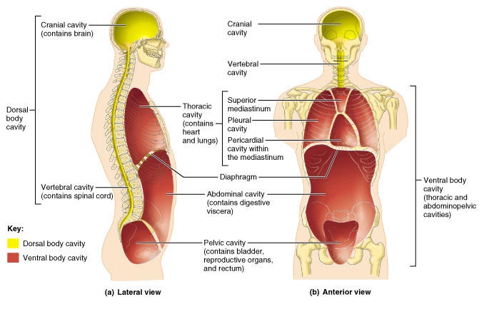 divided into lateral pleural cavities (each pleural cavity envelopes a lung) and the mediastinum. Within The pericardial cavity lies within the mediastinum. It encloses the heart and remaining thoracic organs (trachea, esophagus, ect.).
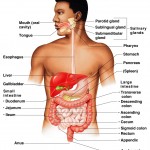
The inferior division of the ventral body cavity is called the “abdominopelvic cavity” and is separated from the thoracic cavity by the diaphragm. The abdominopelvic cavity is also separated into two subdivisions, the “abdominal cavity” and “pelvic cavity“. The abdominal cavity contains the stomach, spleen, liver, intestines, and a few other organs. The pelvic cavity (inferior) contains the urinary bladder, rectum, and some reproductive organs.
Membranes in the Ventral body cavity

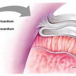
The walls of the ventral body cavity and outer covering of its organs contain a thin covering called the serosa (also called serous membrane). It is a double-layered membrane made up of two parts called the “parietal serosa” (lines the cavity walls) and “visceral serosa” (covers organs in the cavity). The serous membranes are separated by a thin layer of fluid called “serous fluid“. Serous fluid is secreted by both membranes and acts as a lubricant, allowing organs to slide in the cavity without causing friction.
Typically, the serous membranes are named according to the cavity and organ they associate with. For instance, the parietal pericardium lines the pericardial cavity.
Abdominopelvic regions and quadrants”
Because it’s so large, the abdominopelvic cavity is separated into regions and quadrants. The quadrants are self-explanatory and can be figured out fairly easily by looking at the abdominopelvic cavity. They consist of the:
- Right upper quadrant (RUQ)
- Left upper quadrant (LUQ)
- Right lower quadrant (RLQ)
- Left lower quadrant (LLQ)
Simply draw a cross over the cavity seperating it into four boxes, then use the directional terms accordingly.

Abdominopelvic Regions: Image by Mary Weis
The 9 regions of the abdominopelvic cavity are listed below (see image above also).
- Umbilical region– center-most region (belly button)
- Epigastric region– superior to the umbilical region (above belly)
- Hypogastric region– inferior to the umbilical region (pubic area)
- Right and left iliac (inguinal region)-located lateral to the hypogastric region
- Right and left lumbar regions– lateral to the umbilical region
- Right and left hypochondriac regions– lateral to the epigastric region
Other body cavities
- Nasal cavity– is part of the respiratory system. Located within the nose (and posterior).
- Orbital cavities– house the eyes
- Oral cavity– the mouth, contains the teeth and gums
- Synovial cavities– surround freely movable joints and secrete a lubricating fluid like serous membranes.


Category: The Basics


